Smart fabrication techniques

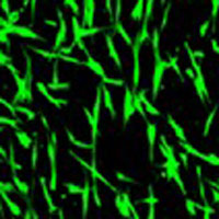
Here in Keele University, we have developed a facile but effective collector with parallel electrodes for low density aligned nanofiber collection in electrospinning technique. The aligned nanofibers demonstrate strong contact guidance for various cellular model establishment. The handleable and portable nanofiber meshes allows to build-up multiple layered 3D tissue models with defined different cellular orientation in the adjacent layers.
Incorporating nanofibers for contact guidance in 3D tissue models

Construction of the orthogonally arranged nanofiber meshes in hydrogel
3D bioprinted organ tissue has the potential for aiding drug screening, formulation development, clinical transplantation, chemical testing, as well as basic research. A key hope for 3D bioprinting tissues is the improved tissue authenticity over conventional engineered constructions, enabling the precise localization of multiple cell types and appendages within a construct. Our team at Keele University has synthesised new bioinks and developed a versatile technique for the fabrication of human skin constructs.

3D bioprinting a full thickness human skin equivalent and characterisation
Electrohydrodynamic atomization (EHDA), also called electrospraying, has been widely employed for encapsulating therapeutic agents in biodegradable polymeric particles and microbubbles for controlled and sustained drug release applications. EHDA produces very fine droplets from a capillary liquid stream by using an electric field. Our team at Keele University has co‐encapsulated chemotherapeutic agents into core‐shell microparticles using C-EHDA technique to locally administer them against tumor cells.

Coaxial Electrohydrodynamic atomization (CEHDA) technique for the production of core-shell structured microparticles

